
Корзина пуста.
12-24 Months,Augma Bone Cement Academy,Bond Apatite®,Bone Cement,Bone Cement Expert,Clinical Cases,Clinical Indication,Clinician,Dental Notation,Images,Lateral Augmentation,Lower Right Molar,Lower Right Premolar,Media,Post-Op Period,Socket Grafting
Lateral Augmentation of Thin Alveolar Ridge – 45 (29) – #47 (31)
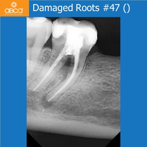
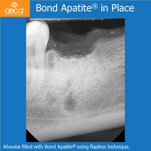
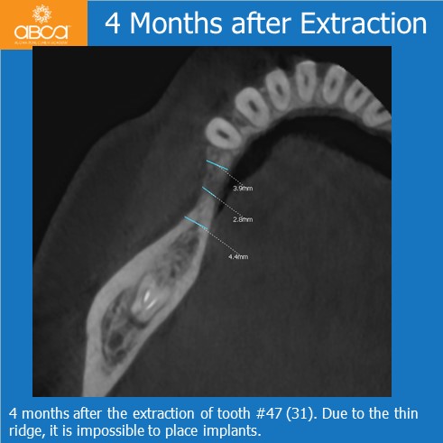
The patient is a healthy, 38 year old female. Reconstruction was planned in the area of #45 (29) – #47 (31). The first step was the extraction of broken tooth #47 (31) due to inflammatory changes in furcation. There was no possibility of prosthetic restoration. The patient had some pain and discomfort. After extraction, the socket is filled with 1 cc Bond Apatite®, without opening a flap. The graft was covered with Augma Shield™. The patient did not follow post-op instructions and chewed hard food above the graft, which caused the graft to fail.
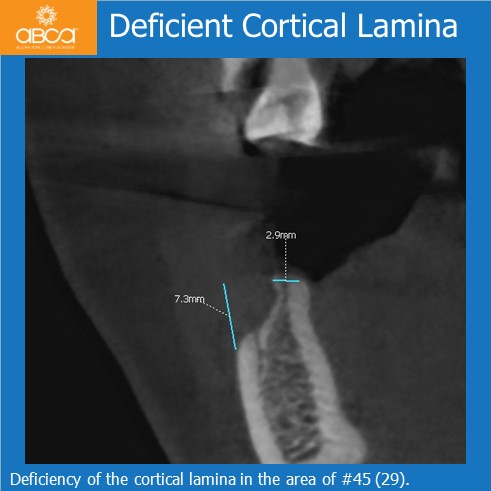
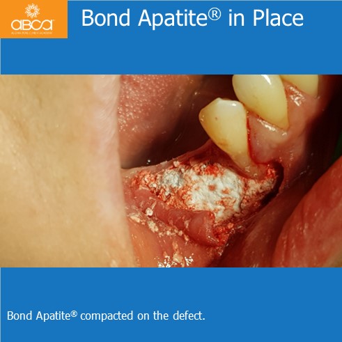
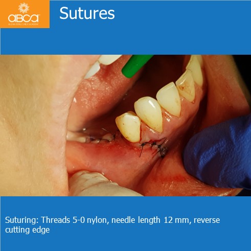
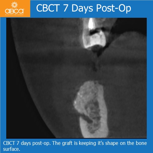
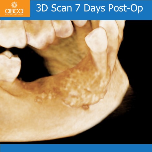
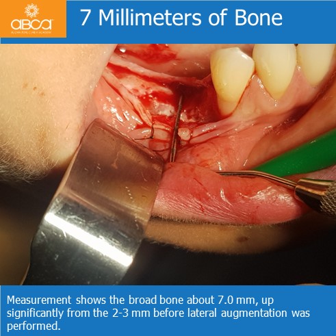
The second step took place 4 months after grafting. A two step alveolar ridge regeneration in area #45 (29) – #46 (30), after which we confirmed a lesion of the external cortical lamina, and measured the difference between both laminas as about 7mm, and the diameter of the top of the ridge medium as 3.0 mm. The bone defect was filled with 2cc Bond Apatite®, with a flap that was closed with tension.
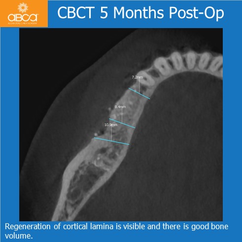
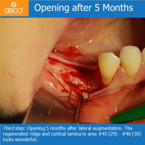
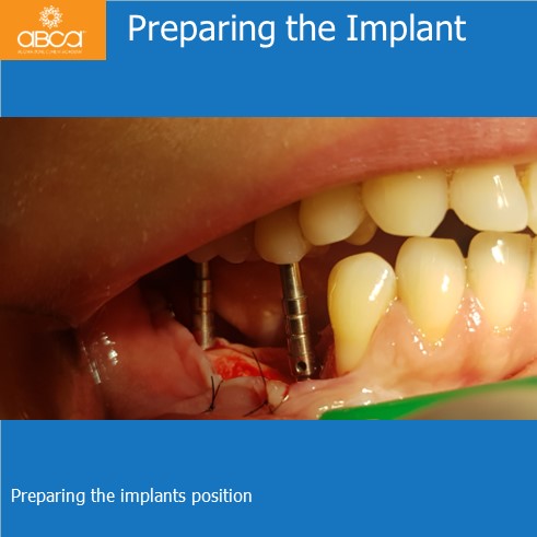
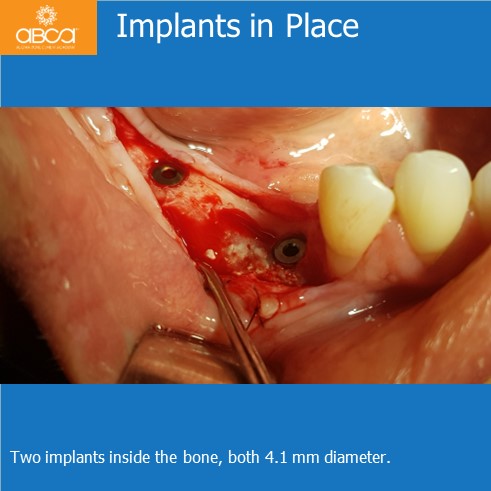
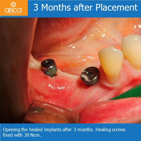
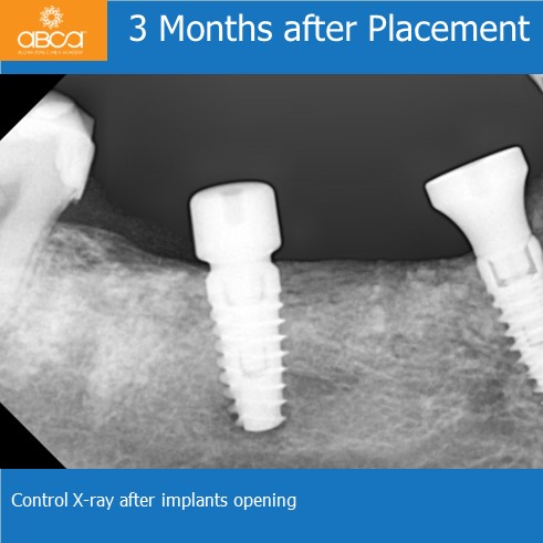
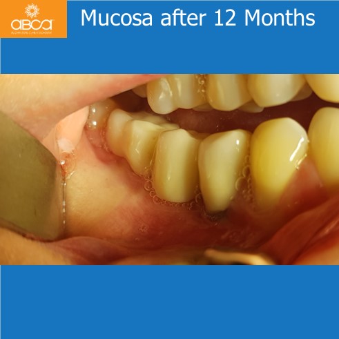
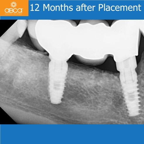
5 months later tomography showed a full restoration of the external lamina, and one month after that two IRES implants were inserted. 4 months after this a final prosthetic restoration was done, and throughout the 12 month follow up no inflammatory symptoms were observed.